Image Gallery
A small collection of illustrative images evoking the world of neuro-oncology — the laboratories, the imaging, and the collaborative spirit that defines the field.
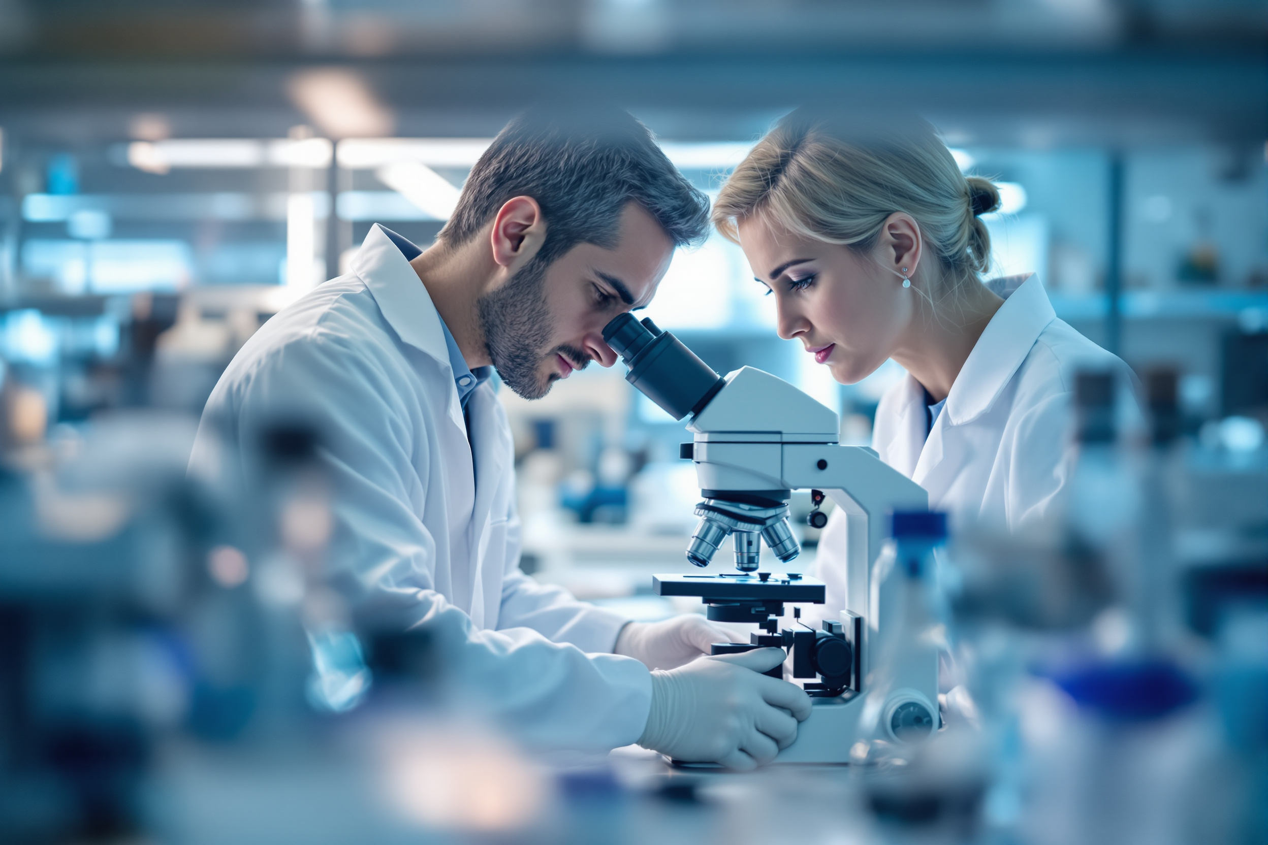
Science and imaging
Modern neuro-oncology lives at the meeting point of the microscope and the scanner. Advances in neuropathology and neuroimaging have transformed how brain tumours are diagnosed, classified, and monitored over time.

The human side
Behind every scan and slide is a person and a family. The collaborative culture documented throughout this archive exists for one reason: to improve outcomes and quality of life for people living with brain tumours. For ways families can find help, see our patient support page.
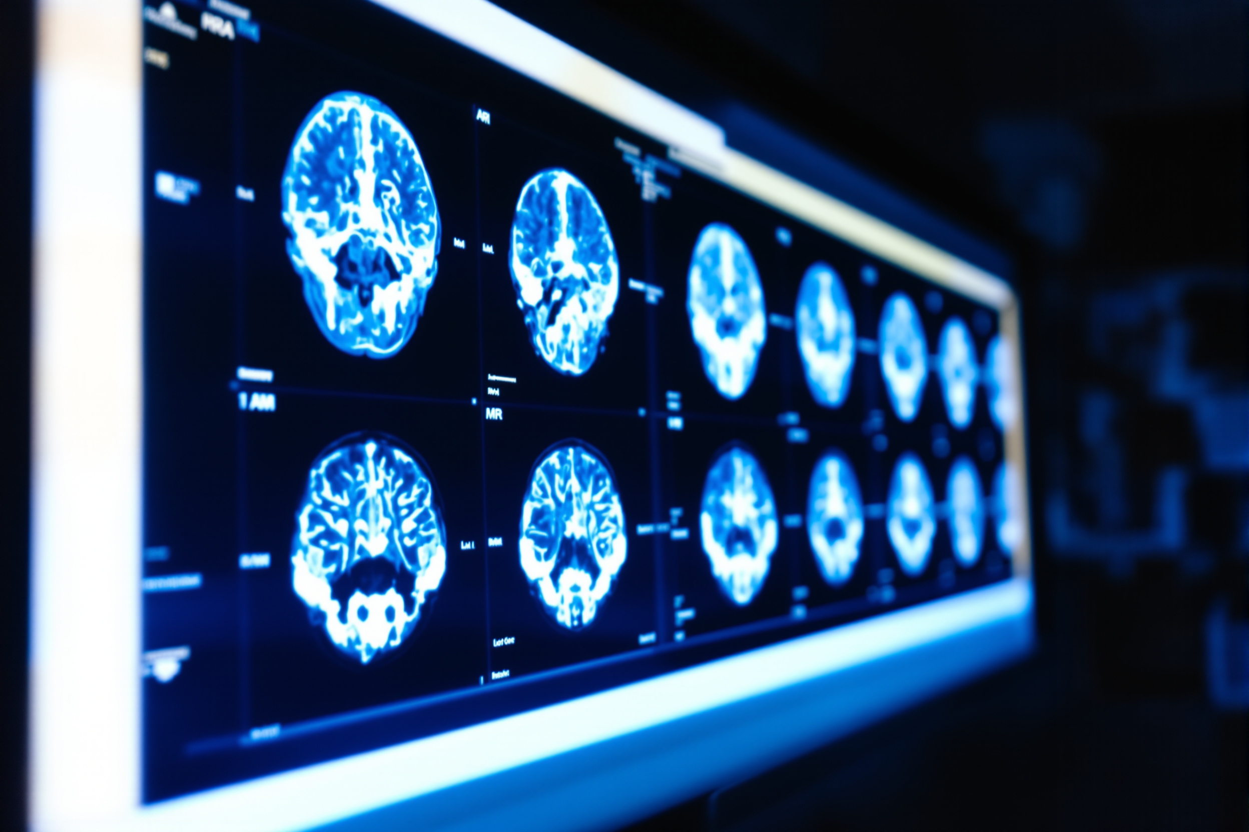
Why imaging defines the field
If any single technology shaped modern neuro-oncology, it is imaging. Magnetic resonance imaging made it possible to see tumours in exquisite detail, plan surgery with precision, and track response to treatment over months and years. Newer techniques — diffusion and perfusion imaging, spectroscopy, and functional mapping — add information about a tumour's biology and its relationship to critical brain regions. Alongside the scanner, the microscope and the molecular laboratory have transformed how tumours are classified, moving the field from appearance alone toward an integrated, biology-informed diagnosis.
A note on imagery
Because authentic meeting photographs feature real individuals and events, we have chosen not to reproduce them. The illustrations on this page are conceptual pictures created for the archive; they are intended only to convey the spirit of the field, not to depict any specific person, institution, or event. To explore the documented history instead, visit the history and the meeting chronology.